STEP2DYNA Newcastle University researchers Stefan Wernitznig and Claire Rind have published a paper titled “The complex synaptic pathways onto a looming-detector neuron revealed using serial block-face scanning electron microscopy” in the Journal of Comparative Neurology. Figure 3 from this paper (as seen below) was featured as the journal’s cover image.
The Journal of Comparative Neurology is a top-tier journal publishing research articles on ultra-structural details of the circuitry revealed using the latest techniques in the central nervous systems of different animal species, including humans. It has a significant impact in understanding function through the detailed structure of pathways for behaviour. These can provide inspiration for scientists wanting to model particular behaviours and mimic their function.
Research Summary
The ability of locusts to detect looming stimuli and avoid collisions or predators depends on a neuronal circuit in the locust’s optic lobe. Although comprehensively studied for over three decades, there are still major questions about the computational steps of this circuit. We used fourth instar larvae of Locusta migratoria to describe the connection between the lobula giant movement detector 1 (LGMD1) neuron in the lobula complex and the upstream neuropil, the medulla. Serial block-face scanning electron microscopy (SBEM) was used to characterize the morphology of the connecting neurons termed trans-medullary afferent (TmA) neurons and their synaptic connectivity. This enabled us to trace neurons over several hundred micrometres between the medulla and the lobula complex while identifying their synapses. We traced two different TmA neurons, each from a different individual, from their synapses with the LGMD in the lobula complex up into the medulla and describe their synaptic relationships. There is not a simple downstream transmission of the signal from a lamina neuron onto these TmA neurons; there is also a feedback loop in place with TmA neurons making outputs as well as receiving inputs. More than one type of neuron shapes the signal of the TmA neurons in the medulla. We found both columnar and trans-columnar neurons connected with the traced TmA neurons in the medulla. These findings indicate that there are computational steps in the medulla that have not been included in models of the neuronal pathway for looming detection.
Technical summary
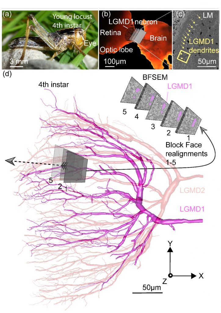
Figure 1 demonstrates the work-flow showing critical points, from locating the lobula giant movement detector 1 neuron (LGMD1) in the lobula region, to following the neurons synapsing with the LGMD1 back to their origin in the medulla.
(a) Fourth instar of the locust Locusta migratoria.
(b) Initial block orientation to locate the LGMD1 shown in reference to light microscope image of an adult optic lobe in which the LGMD1 had previously been stained intracellularly.
(c) Block face of the outer lobula region (OLO) in the optic lobe of a fourth instar locust in sagittal orientation. The crescent of LGMD1 and 2 branches are visible in cross section in the OLO (LGMD1 marked with arrows). A yellow rectangle indicates the size of the cut-down block-face used to reconstruct neurons using SEM and is the starting point for the tracing of TmAs synaptically linked to the LGMD1.
(d) Serial block-face scanning electron microscopy (SBEM) images of block-faces taken during TmA tracing, superimposed of reconstruction of silver stained LGMD neurons (subfield A) in the optic lobe of a fourth instar locust (method as in Sztarker & Rind, 2014). Continual manual block realignments were necessary to follow the TmAs into the medulla (adjustments 1–5 shown).
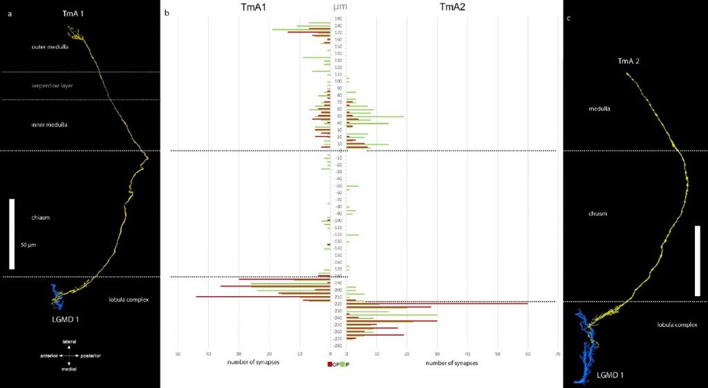
LGMD1 inputs are not simply relaying-on the information they receive. In Figure 2, two inputs to the LGMD1 are reconstructed to the same scale. The TmAs (in yellow on black panels labelled a and c) synapse and provide input to the LGMD1 (blue). TmAs have inputs (b, shown in green) and make outputs (b,shown in red) in the medulla. The medulla and lobula regions of the optic lobe are separated by the chiasm a region with few synapses. TmA1 and 2 were mapped in different locusts.
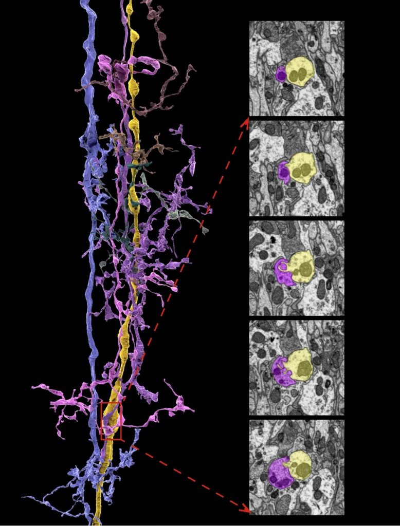
Figure 3 demonstrates TmA neurons, coming from the eye and that later provides input to the LGMD1, also synapse with a range of neurons earlier in the medulla. The TmA2 is shown here on the left and right panels in yellow and the synapsing neurons are shown in blues, magenta and purple. The TmA2 neuron that contacts the LGMD1 has very short branches in the medulla where it makes connections suggesting it could transmit information quickly, because signals travel more slowly in small diameter branches. The TmA2 has swellings where small beads with out-pushings make and receive synapses (right). We have described complex circuitry, involving local processing on the way to the LGMD1 that could be used to improve the performance of models of collision detection by the LGMD. Physiological experiments could provide details of the dynamic features of the interactions.
The full paper can be found as open access on the Wiley Online Library and on the UNEW Eprints Portal.